Pandemic – COVID-19: The Coronavirus Disease Uncovered

The Coronavirus is a pathogen that has a direct impact on the respiratory system. In the past, this type of virus had caused other outbreaks; namely the severe acute respiratory syndrome SARS and Middle East Respiratory Syndrome MERS. COVID-19 is a potentially fatal disease caused by SARS-CoV2 (Novel Coronavirus) (Watts et. al, 2020). This strain is of great concern amongst health officials due to its high R0 value. The R0 estimates ranged from 2.06-2.52, which indicates the pathogen’s great infectivity.
• If R0 is less than 1, each existing infection causes less than one new infection. In this case, the disease will decline and eventually die out.
• If R0 equals 1, each existing infection causes one new infection. The disease will stay alive and stable, but there will not be an outbreak or an epidemic.
• If R0 is more than 1, each existing infection causes more than one new infection. The disease will spread between people, and there may be an outbreak or epidemic.
Table 1. R0 values and their implications.
The initial cluster of patients, identified in December 2019, upon admission to the hospitals were initially diagnosed with pneumonia of unknown aetiology. Based on epidemiological evidence this cluster was linked to the Hua Nan seafood and live animal market in Wuhan, Hubei Province, China (Watts et.al, 2020).
The patients’ preliminary symptoms included fever, cough, tiredness and dyspnoea (shortness of breath). The antecedent clinical manifestations of COVID-19 can be classified into two categories; namely systemic and respiratory disorders. The systemic disorders are sputum production, haemoptysis which involves instances of coughing up blood or blood-stained mucous, acute cardiac injury, hypoxemia (low oxygen levels in the blood mainly in the arteries), lymphopenia (reduced lymphocyte count) and diarrhoea. The respiratory disorders include sneezing, sore throat, rhinorrhea, pneumonia and acute respiratory distress syndrome. (Huang, et. al, 2020).
The pathogen’s high infectivity made it imperative for early testing and diagnosis to be conducted. The preliminary testing methods used for diagnosis are body temperature scans, chest CT scans, X-rays and blood work. Confirmatory tests involve specimen collection; nasopharyngeal (NP) swab are preferred although an oropharyngeal swab is also acceptable. These specimens are then tested using serological methods, viral sequencing and most importantly nucleic acid amplification tests (NAAT) for COVID-19.

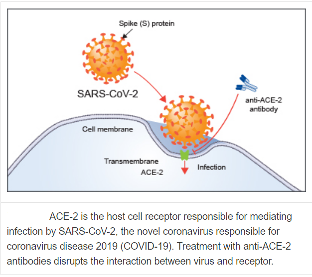
How exactly does the SARS-CoV2 manage to cause such life-threatening conditions? The virus makes its way into your body via the droplets of an infected person. These droplets are released by coughing, sneezing and even when breathing. The virus could travel from the infected person through the air or land on surfaces and enter a healthy person via the mouth, nose or the eyes through touch. This provides the virus with a pathway via the mucous membrane. Upon entry, the virus will bind to the ACE2 receptors present on the lungs’ epithelial cells, thus hijacking the healthy cells and eventually destroying them. (Hussin et. al, 2020)
Patients who test positive for COVID-19, exhibit abnormal respiratory symptoms along with elevated pro-inflammatory cytokines (chemical messenger for immune cells). Some other laboratory findings include increased red blood cell sedimentation rate, leukopenia (decrease of white blood cells) of which 70% are neutrophils. (Huang et. al, 2020). Neutrophils are significant as they are the front liners in our immune systems response. Additionally, there are high levels of blood C-reactive protein which is an indicator that the immune system is active and responding to inflammation. The cells in the immune system will also secrete cytokines who act as chemical messengers e.g. TNFα. When the patients’ chest CT scans are taken, they reveal pneumonia, RNAaemia (detection of cytomegalovirus RNA i.e genetic material in the serum) coupled with ground-glass opacities (hazy opacities in CT scans that indicate filled air spaces in the lungs due to exudate accumulation).

At present, there are no vaccines or antiviral therapies available for COVID-19. The current treatment programme includes the use of broad-spectrum antiviral medications such as oseltamivir, lopinavir and ritonavir. The National Institute of Health is currently conducting trials to test hydroxychloroquine as a potential treatment option for COVID-19. (National Institute of Health, 2020) Current research is now focused on understanding the biology of the virus, potential treatment option and most importantly the psychological effects that a pandemic like situation has on the society along with the socio-economic changes.
During such a pandemic, our best defence is to maintain good personal hygiene, practise social distancing and strictly follow the guidelines provided by government healthcare officials.
References
- Bogoch, A. Watts, A. Thomas-Bachli, C. Huber, M.U.G. Kraemer, K. Khan
Pneumonia of unknown etiology in Wuhan, China: potential for international spread via commercial air travel. J. Trav. Med. (2020).
- C. Huang, Y. Wang, X. Li, L. Ren, J. Zhao, Y. Hu, et al. Clinical features of patients infected with 2019 novel coronavirus in Wuhan, China Lancet, 395 (10223) (2020).
- Hussin A.Rothana. Siddappa N.Byrareddy, The epidemiology and pathogenesis of coronavirus disease (COVID-19) outbreak. Elsevier, Journal of Autoimmunity Feb 26 2020.
- Prof Chaolin Huang, MD, Yeming Wang, MD, Prof Xingwang Li, MD, Prof Lili Ren, PhD, Prof Jianping Zhao, MD, Yi Hu, MD, Clinical features of patients infected with 2019 novel coronavirus in Wuhan, China. The Lancet, Volume 395, Issue 10223, 497 – 506.
- Laboratory testing for Middle East Respiratory Syndrome coronavirus, interim guidance (revised), January 2019, WHO/MERS/LAB/15.1/Rev1/2019, World Health Organization, 2018.
- Zhang S, Diao M, Yu W, Pei L, Lin Z, Chen D. Estimation of the reproductive number of novel coronavirus (COVID-19) and the probable outbreak size on the Diamond Princess cruise ship: A data-driven analysis. Int J Infect Dis. 2020; 93: 201–204.
This article is contributed by Norishka Cassy Noel D’lima from MDIS School of Life Sciences.


