MDIS School of Life Sciences Immersion Programme

MDIS School of Life Sciences (SLS) conducted the immersion programme for students from Korea Bio Meister High School. This would be the second consecutive year of the immersion programme.
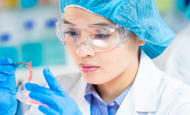
Throughout the immersion programme, students were introduced to various laboratory techniques and also the instruments in the MDIS School of Life Sciences laboratories. The students paired up for short projects to be completed within a span of six days. The students were closely supervised and guided for the optimum laboratory experience as they learnt more on the below techniques and procedures.
Microbiological techniques
Microbiological techniques are methods used for the study of microbes, including bacteria and microscopic fungi.
The immersion programme students had the opportunity to perform these techniques to survey, culture, stain, identify, engineer and manipulate microbes. Pour plate, serial dilution, biochemical tests and bacterial staining techniques were some of the techniques introduced to the students.
Laminar air flow cabinet
One of the highlights during the programme was working in the laminar air flow cabinet. This special cabinet allows their agar media plate experiments to be carried out in a controlled and uncontaminated environment, for more accurate results.
Students prepared their own agar media and sterilised them using the autoclave. The autoclave is important for all microbiological experiments, as it provides a physical method for sterilisation of all the media and components required for their experiments.
Identification of bacteria
Students were trained to use the API (Analytical Profile Index) test system, which facilitates faster identification of bacteria. These test kits provide manual identification of many industry-relevant microorganisms, as well as infectious disease-causing microorganisms.
The students also went through extensive observation of the microbial samples under the microscopes. They were then taught the Gram staining technique to differentiate between gram positive and gram negative bacteria, where the bacteria would pick up either a violet/purple (Gram positive) or red/pink (Gram negative) coloured stain.
Through this process, the students were able to view Escherichia coli, Staphylococcus species, Streptococcus species, and spore forming bacteria like the Bacillus species.
At the end of the six-day immersion programme, the students had to recap and collate all their findings throughout. The students did a commendable job in presenting their scientific findings throughout the experiments conducted during the six-day programme, in the form of graphs and tables.
Six days may have been a short period of time to master the lab procedures. Nonetheless, it was a fruitful experience for them as the students walked away from the immersion programme with valuable insights, while getting a taste of what the MDIS School of Life Sciences entails.
The article is contributed by Dr Sunesh Keecheril Augustine, Senior Lecturer, MDIS School of Life Sciences
References:
Cappuccino, J. G., & Sherman, N. (2014). Microbiology: a laboratory manual. Boston, Pearson Education.
Collins, C. H., Lyne, P. M., & Grange, J. M. (1987). Collins and Lyne’s Microbiological methods. London: Butterworths.
Goering, R. V., & Mims, C. A. (2008). Mims’ Medical microbiology. Philadelphia, PA: Mosby Elsevier.
Madigan, M. T., Martinko, J. M., & Brock, T. D. (2006). Brock biology of microorganisms. Upper Saddle River, NJ: Pearson Prentice Hall.
Truant, A. L. (2016). Manual of Commercial Methods in Clinical Microbiology. New Jersey: John Wiley & Sons Inc.


